Post-doc / Faculty Seminar Series
Date: June 18, 2015
Location: K-307 (3-6408), University of Rochester Medical Center
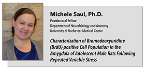
Characterization of Bromodeoxyuridine (BrdU)-positive Cell Population in the Amygdala of Adolescent Male Rats Following Repeated Variable Stress
Michele Saul, Ph.D.
Postdoctoral Associate, Fudge Lab
Department of Neurobiology and Anatomy
University of Rochester Medical Center
Adolescence is a developmental stage characterized by changes in neural organization. During this period, the amygdala, a structure in the temporal lobe that mediates emotion and social behaviors, is changing in terms of neural and glia density. My research focuses on determining how the amygdala changes during normal adolescent development, and how this trajectory can be altered by the addition of a stressful event in early adolescence.
Cell proliferation is one type of plasticity that is affected by development and stress in many brain regions. In the amygdala, we found that the number of bromodeoxyuridine (BrdU; a marker of DNA replication) positive cells in adolescent rats is 4-5 times higher than in young adult animals. This suggests that adolescence is a relatively active period of cell proliferation and plasticity in this region, compared to young adulthood. BrdU pulse-chase studies indicated that at least a portion of cells divide locally in the adolescent amygdala, leaving open the possibility that dividing cells are also migrating in. Further characterization with neural markers showed that approximately 30% of BrdU-positive cells co-localize with doublecortin (DCX; a putative marker of immature neurons), but the cells do not show the developmental profile of typical neuronal precursors. The BrdU-positive population had a 50% co-localization with neural-glial antigen (NG2), which identifies a unique type of oligodendrocyte precursor.
To determine whether stress influences cell proliferation in early adolescence, a repeated variable stress (RVS) paradigm was used to determine how stress influences BrdU-positive cells in the adolescent amygdala. Adolescent rats were administered RVS (post natal day (PND) 27-29), injected with BrdU (PND 30-31), and sacrificed either 24 hours (PND 32) or 10 days later (PND 41). In stressed animals, the number of BrdU-positive cells significantly decreased 10 days post-BrdU but did not change 24 hours post-BrdU. This suggests that RVS exerts effects over a delayed time course, possibly through apoptosis or cell cycle arrest. Characterization of the BrdU-positive cells present at the 10-day chase time point showed that the number of DCX/BrdU double-labeled cells were not altered by stress, but the number of NG2/BrdU double-labeled cells was significantly decreased by stress.
Taken together, these studies suggest that adolescence is an active time for amygdala development and cell proliferation. Although many cells contain DCX, they do not mature into neurons in the typical time course. Therefore, they might be late-developing neurons or might not be neurons at all. DCX-positive cells may also represent a subpopulation of NG2 cells, which was not examined. Many BrdU-labeled cells were NG2-positive, which suggests that they may be oligodendrocyte precursor cells. Stress significantly influences the NG2/BrdU population, which suggests that there is a significant effect of stress on gliogenesis during adolescence. These changes in gliogenesis may lead to long-lasting effects for the animal, including alterations in network functionality, changes in behavior and reactivity to future stress exposure.
Refreshments will be provided
Sponsored by the Rochester Chapter of the Society for Neuroscience
Stay Up-To-Date
Subscribe to the SFN Rochester Chapter LISTSERV to receive news and events via email.

